Beyond TSH: Why Your Thyroid Might Still Be the Problem

Quick Answer
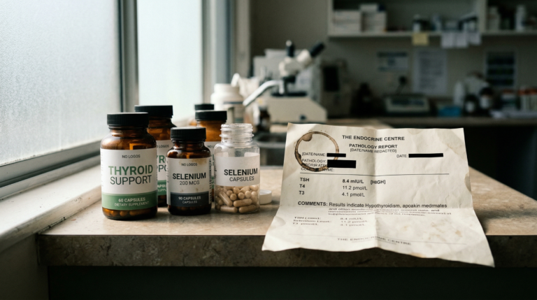
TSH (thyroid-stimulating hormone) alone may miss significant thyroid dysfunction. This pituitary-derived marker does not reflect peripheral T4-to-T3 conversion, cellular hormone uptake, or autoimmune thyroid activity such as Hashimoto’s thyroiditis. A comprehensive thyroid panel including free T4, free T3, reverse T3, and thyroid peroxidase antibodies (TPOAb) can reveal subclinical dysfunction that standard TSH screening often overlooks.
In this article, we explore why TSH alone may miss underlying issues and what deeper testing can reveal about your thyroid health.
At a Glance
- TSH reflects pituitary signalling only and may not detect impaired T4-to-T3 conversion in peripheral tissues
- Hashimoto’s thyroiditis accounts for approximately 70-90% of hypothyroidism cases in iodine-sufficient regions and can be present years before TSH becomes abnormal
- Selenium-dependent deiodinase enzymes (D1, D2, D3) are required for converting inactive T4 into biologically active T3
- Elevated reverse T3 (rT3) may reduce effective thyroid hormone signalling during periods of chronic stress or inflammation
- Nutrient cofactors including iodine, selenium, iron, zinc, and vitamin D are essential for thyroid hormone synthesis and receptor binding
- Research by Wartofsky and Dickey (2005) suggests the TSH reference range may be too broad to detect subclinical hypothyroidism in symptomatic individuals
Why TSH Alone Is Not Enough
TSH (thyroid-stimulating hormone), produced by the anterior pituitary gland, serves as a feedback signal to the thyroid gland via the hypothalamic-pituitary-thyroid (HPT) axis. While TSH is a useful screening marker, it primarily reflects pituitary signalling rather than downstream thyroid hormone availability or action at the tissue and cellular level (4,10). As Hoermann et al. demonstrated in their 2015 analysis of thyroid-pituitary homeostasis, TSH and peripheral thyroid hormones do not always move in predictable lockstep.
A normal TSH does not indicate:
- Whether sufficient thyroid hormone is being produced
- Whether T4 is being effectively converted into active T3 by deiodinase enzymes
- Whether thyroid hormones are entering cells efficiently via monocarboxylate transporter 8 (MCT8)
- Whether autoimmune processes are impairing thyroid tissue
As a result, individuals may experience classic hypothyroid symptoms despite a TSH value that falls within laboratory reference ranges (5,6).
What Full Thyroid Testing Includes
A comprehensive thyroid assessment measures multiple biomarkers across the HPT axis, each reflecting a distinct aspect of thyroid function.
| Marker | What It Measures | Clinical Relevance |
|---|---|---|
| TSH | Pituitary signalling to the thyroid | Screening marker; does not reflect peripheral hormone action |
| Free T4 (thyroxine) | Primary circulating storage hormone | Indicates thyroid gland output |
| Free T3 (triiodothyronine) | Biologically active hormone at the cellular level | Reflects effective thyroid activity in tissues |
| Reverse T3 (rT3) | Inactive metabolite of T4 | May competitively inhibit T3 action at thyroid receptors |
| TPOAb (thyroid peroxidase antibodies) | Autoimmune marker targeting thyroid peroxidase enzyme | Elevated in Hashimoto’s thyroiditis (1-3) |
| TgAb (thyroglobulin antibodies) | Autoimmune marker targeting thyroglobulin protein | Additional autoimmune thyroid marker (1-3) |
This type of assessment is commonly used in functional and integrative approaches to thyroid dysfunction, where symptom persistence is explored beyond basic screening markers.
T4-T3 Conversion
Approximately 80% of circulating T3 is produced outside the thyroid gland through peripheral conversion of T4, primarily by selenium-dependent deiodinase enzymes (type 1 and type 2 deiodinase, known as D1 and D2) in the liver, kidneys, and skeletal muscle (7,8). Factors that may impair this conversion include:
- Chronic psychological or physiological stress and elevated cortisol
- Systemic inflammation and elevated inflammatory cytokines (IL-6, TNF-alpha)
- Selenium, zinc, or iron deficiency
- Hepatic dysfunction affecting liver-based T4-to-T3 conversion
- Acute or chronic illness (non-thyroidal illness syndrome / euthyroid sick syndrome)
When peripheral conversion is impaired, functional thyroid activity may be reduced despite normal TSH and T4 levels (7-10). Bianco et al. (2019) provided a comprehensive review of the cellular and molecular biology underlying these conversion pathways.
Reverse T3
Reverse T3 is an inactive isomer of T3 produced preferentially by type 3 deiodinase (D3) during periods of physiological stress, caloric restriction, illness, or inflammation (9,10). Chronically elevated rT3 may reduce effective thyroid hormone signalling at the cellular level by competing with T3 for binding at thyroid hormone nuclear receptors (thyroid receptor alpha and beta). Its clinical relevance remains debated within endocrinology, which is why interpretation should always be cautious and contextual.
The Autoimmune Connection
Hashimoto’s thyroiditis, first described by Japanese physician Hakaru Hashimoto in 1912, accounts for approximately 70-90% of hypothyroidism cases in iodine-sufficient regions such as Australia, according to research by McLeod and Cooper (2012) and Vanderpump (2011) (1,2). Importantly, autoimmune activity marked by elevated TPOAb and TgAb can be present years before measurable changes in TSH occur (3,11).
Testing thyroid antibodies may therefore be clinically relevant in symptomatic individuals even when TSH appears normal, recognising that autoimmune progression varies substantially between individuals. Autoimmune thyroid disease is also frequently associated with broader gut-immune interactions, including intestinal permeability and molecular mimicry, which can influence inflammatory load and immune signalling.
The Nutrient Factor
Thyroid hormone synthesis, activation, and receptor signalling depend on adequate availability of several key micronutrients, each playing distinct roles in the HPT axis.
| Nutrient | Role in Thyroid Function | Clinical Consideration |
|---|---|---|
| Iodine | Required substrate for T4 and T3 synthesis by thyroid peroxidase | Excess may exacerbate autoimmunity; testing recommended before supplementation (12) |
| Selenium | Cofactor for deiodinase enzymes (D1, D2) and glutathione peroxidase antioxidant protection | Kohrle (2005) demonstrated selenium’s role in thyroid hormone metabolism (11) |
| Iron (ferritin) | Required for thyroid peroxidase enzyme activity | Iron deficiency associated with impaired thyroid hormone action (15) |
| Zinc | Supports thyroid hormone receptor binding | Deficiency may reduce T3 production and receptor sensitivity (11-13) |
| Vitamin D | Immune regulation and modulation of autoimmune thyroid activity | Low vitamin D status associated with higher TPOAb levels (11-13) |
| Vitamin A | Thyroid hormone receptor binding and gene transcription | Works synergistically with thyroid hormones at the nuclear receptor level (11-13) |
Because both deficiency and excess can be harmful, testing before supplementation is clinically important, particularly in autoimmune thyroid disease (11-13).
Why Functional Ranges Matter
Laboratory reference ranges are statistically derived from population samples and designed to identify overt disease rather than optimise symptom resolution. Wartofsky and Dickey’s influential 2005 paper in the Journal of Clinical Endocrinology and Metabolism argued for a narrower thyrotropin reference range, suggesting that many individuals with TSH values in the upper reference range may still experience subclinical hypothyroidism (4). Andersen et al. (2002) further demonstrated that individual TSH set points vary considerably and are much narrower than population-based reference ranges.
For example:
- TSH values in the upper reference range (e.g., 3.0-4.5 mIU/L) may still be associated with hypothyroid symptoms in some individuals (4,6)
- Low-normal free T3 has been linked with fatigue, weight gain, and cognitive symptoms in some individuals (5,7)
Clinical interpretation should therefore integrate biochemical data with symptoms and individual context, rather than relying on reference ranges alone (6,10).
Thyroid Medication Isn’t Always the Whole Answer
Levothyroxine (synthetic T4) remains the standard of care for hypothyroidism and is essential and life-changing for many people (14,15). However, when thyroid dysfunction is driven or compounded by stress physiology, systemic inflammation, nutrient deficiencies, or autoimmune activity, medication alone may not fully resolve symptoms (9,13,15). Chaker et al. (2017) noted in their Lancet review that approximately 5-10% of levothyroxine-treated patients remain symptomatic despite normalised TSH.
Some clinicians consider combination therapy (T4 plus T3) or desiccated thyroid extract in selected cases, though evidence remains mixed. Medication requirements vary between individuals and should only be adjusted under appropriate medical supervision (14,15).
The Stress-Thyroid Relationship
Chronic hypothalamic-pituitary-adrenal (HPA) axis activation and elevated cortisol are associated with measurable changes in thyroid function, including:
- Reduced T4-to-T3 conversion via suppression of type 2 deiodinase (D2) activity
- Increased reverse T3 production via upregulation of type 3 deiodinase (D3)
- Altered hypothalamic-pituitary-thyroid (HPT) axis signalling, including suppressed thyrotropin-releasing hormone (TRH) output (9,13)
Persistent stress physiology is also a common contributor to chronic fatigue presentations, particularly when thyroid markers appear “normal” on standard testing. Fliers et al. (2015) explored these mechanisms in their review of thyroid function in critically ill patients, demonstrating how non-thyroidal illness syndrome can mimic hypothyroidism biochemically.
When to Consider Comprehensive Thyroid Testing
Comprehensive thyroid testing may be worth considering if you:
- Have hypothyroid symptoms (fatigue, cold intolerance, constipation, dry skin) despite a normal TSH
- Have a family history of autoimmune thyroid disease or other autoimmune conditions (coeliac disease, type 1 diabetes, rheumatoid arthritis)
- Are taking levothyroxine or other thyroid medication but remain symptomatic
- Experience unexplained fatigue, weight resistance, hair loss, or brain fog (5,6,15)
Next Steps
- Request a full thyroid panel: Ask for TSH, free T4, free T3, reverse T3, and thyroid antibodies (TPOAb, TgAb) to get a complete picture of thyroid function beyond standard screening.
- Assess nutrient cofactors: Test iodine, selenium, iron (ferritin), zinc, vitamin A, and vitamin D, all of which are required for thyroid hormone synthesis and conversion.
- Evaluate stress physiology: If chronic stress, fatigue, or cortisol dysregulation are present, these factors may be impairing thyroid conversion and should be addressed alongside thyroid management.
Frequently Asked Questions
Key Insights
- A normal TSH does not exclude thyroid dysfunction, it reflects pituitary signalling only
- Hashimoto’s thyroiditis (autoimmune thyroid disease) often precedes abnormal TSH by years
- T3 availability and cellular utilisation via deiodinase enzymes and MCT8 transporters matter clinically
- Nutrient status (selenium, iodine, iron, zinc, vitamin D) and stress physiology significantly influence thyroid function
- Individualised interpretation using functional ranges is essential for symptom resolution
Citable Takeaways
- Hashimoto’s thyroiditis accounts for approximately 70-90% of hypothyroidism cases in iodine-sufficient regions, and autoimmune activity can be present years before TSH becomes abnormal (McLeod and Cooper, 2012; Vanderpump, 2011).
- Approximately 80% of circulating T3 is produced outside the thyroid gland through peripheral conversion of T4 by selenium-dependent deiodinase enzymes in the liver and kidneys (Bianco et al., 2019).
- Wartofsky and Dickey (2005) proposed a narrower TSH reference range, suggesting that upper-range TSH values may still be associated with subclinical hypothyroidism in symptomatic individuals.
- Approximately 5-10% of levothyroxine-treated hypothyroid patients may remain symptomatic despite normalised TSH levels, suggesting additional factors beyond hormone replacement are involved (Chaker et al., 2017).
- Selenium serves as a required cofactor for deiodinase enzymes (D1, D2) responsible for T4-to-T3 conversion and for glutathione peroxidase, which provides antioxidant protection to thyroid tissue (Kohrle, 2005).
- Chronic HPA axis activation and elevated cortisol are associated with reduced T4-to-T3 conversion and increased reverse T3 production via upregulation of type 3 deiodinase (Fliers et al., 2015).
Move Beyond TSH, Get Real Answers About Your Thyroid
If you’ve been told your thyroid is “normal” but symptoms persist, a more complete assessment may help clarify what’s happening. At Elemental Health and Nutrition, Rohan Smith uses comprehensive thyroid panels and functional interpretation to uncover the conversion, autoimmune, and nutrient factors that standard testing often misses.
References
- McLeod DS, Cooper DS. The incidence and prevalence of thyroid autoimmunity. Endocrine. 2012.
- Vanderpump MPJ. The epidemiology of thyroid disease. Br Med Bull. 2011.
- Weetman AP. Autoimmune thyroid disease. Autoimmunity. 2004.
- Wartofsky L, Dickey RA. The evidence for a narrower thyrotropin reference range. J Clin Endocrinol Metab. 2005.
- Andersen S, et al. Narrow individual variations in thyroid function. J Clin Endocrinol Metab. 2002.
- Peterson SJ, et al. An online survey of hypothyroid patients. J Clin Endocrinol Metab. 2018.
- Bianco AC, et al. Biochemistry, cellular and molecular biology of thyroid hormone action. Endocr Rev. 2019.
- Escobar-Morreale HF, et al. Serum thyroid hormone content. Clin Endocrinol. 2005.
- Fliers E, et al. Thyroid function in critically ill patients. Lancet Diabetes Endocrinol. 2015.
- Hoermann R, et al. Homeostatic control of the thyroid-pituitary axis. Front Endocrinol. 2015.
- Kohrle J. Selenium and thyroid hormone metabolism. Thyroid. 2005.
- Zimmermann MB. Iodine deficiency and thyroid disorders. Endocr Rev. 2009.
- Wiersinga WM. Stress and thyroid autoimmunity. J Endocrinol Invest. 2011.
- Boelaert K, Franklyn JA. Thyroid hormone replacement therapy. Lancet. 2005.
- Chaker L, et al. Hypothyroidism. Lancet. 2017.