Mycotox Mould Testing Adelaide: Hidden Drivers of Fatigue

Quick Answer
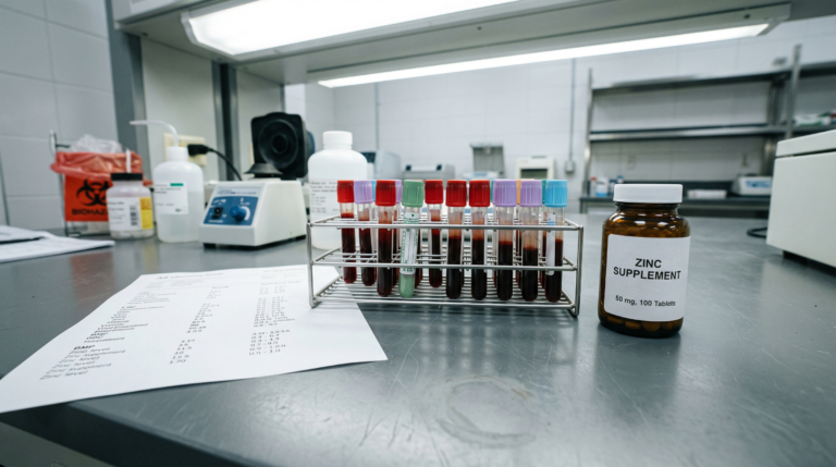
Mycotoxin urine testing, analysed via LC-MS/MS (liquid chromatography-mass spectrometry), detects metabolites produced by indoor moulds including Aspergillus, Penicillium, and Stachybotrys chartarum. Unlike standard IgE mould allergy panels, this clinical assessment measures the body’s current toxic burden by identifying compounds such as ochratoxin A, aflatoxins, and trichothecenes being actively excreted. Research associates these mycotoxins with inflammatory cascades, mitochondrial stress, and neurological symptoms in genetically susceptible individuals (1,2,3,4,10,15).
At a Glance
- Mycotoxins are lipophilic secondary metabolites produced by Aspergillus, Penicillium, Stachybotrys, and Fusarium species commonly found in water-damaged buildings (1,6).
- Urine LC-MS/MS panels can detect ochratoxin A, aflatoxins, trichothecenes, and gliotoxins at sub-nanogram concentrations (2,3,9).
- Ochratoxin A and aflatoxin B1 may impair electron transport chain complexes, reducing ATP synthesis and contributing to chronic fatigue (4,6).
- Glutathione depletion from ongoing mycotoxin exposure may increase oxidative stress and contribute to brain fog and cognitive symptoms (7,12).
- Functional medicine detoxification support may include activated charcoal binders, liposomal glutathione, and methylation cofactors such as methylfolate and methylcobalamin (8,10,11).
For many people in Adelaide, a seemingly healthy home may conceal a silent physiological stressor. Water-damaged buildings (WDB) can harbour indoor moulds that produce mycotoxins, biologically active secondary metabolites that may contribute to chronic inflammatory response syndrome (CIRS) and impaired cellular energy production. Mycotox mould testing Adelaide helps detect harmful mould exposure and mycotoxins that can contribute to chronic fatigue, respiratory issues, and overall detoxification challenges, offering a crucial step in supporting recovery and health optimisation. At Elemental Health and Nutrition, Rohan Smith (BHSc Nutritional Medicine) uses advanced mycotoxin assessment to help identify these potential contributors in individuals experiencing chronic fatigue and post-viral illness.
The Science: How Mycotoxins May Affect Energy Production
Mycotoxins are typically lipophilic compounds that can cross cell membranes and accumulate in fatty tissues such as the brain, liver, and adipose tissue. Research by Bennett and Klich (2003) and Meissonnier et al. (2008) has described several mechanisms through which these toxins may impair cellular function:
| Mechanism | Key Mycotoxins Involved | Potential Effect |
|---|---|---|
| Mitochondrial dysfunction | Ochratoxin A (OTA), aflatoxin B1 | May impair electron transport chain complexes I and III, reducing adenosine triphosphate (ATP) production and contributing to fatigue (4,6) |
| Glutathione depletion | Trichothecenes, deoxynivalenol (DON) | Detoxification relies on glutathione (GSH), the body’s primary intracellular antioxidant; ongoing exposure may increase reactive oxygen species (ROS) and contribute to brain fog (7,12) |
| Immune dysregulation | Gliotoxin, T-2 toxin | May suppress natural killer (NK) cell activity and shift Th1/Th2 cytokine balance, potentially affecting immune resilience and viral control (5,13) |
Why Urine Testing Is Used in Chronic Illness Assessment
LC-MS/MS (liquid chromatography-tandem mass spectrometry) urine mycotoxin panels are considered a reference-standard analytical method for assessing internal mycotoxin exposure in clinical practice. Crinnion (2012) documented the clinical utility of this approach, which is valued because:
| Advantage | Details |
|---|---|
| Direct measurement | Reflects mycotoxins the body is actively eliminating via Phase II conjugation pathways, rather than environmental presence alone (2,11) |
| Broad screening | Panels may include trichothecenes (including deoxynivalenol), gliotoxins, ochratoxin A (OTA), and aflatoxins B1/B2/G1/G2, commonly associated with WDB exposure (1,14) |
| Analytical sensitivity | LC-MS/MS techniques can detect sub-nanogram concentrations, offering greater analytical sensitivity than ERMI or environmental air sampling alone (3,9) |
At Elemental Health and Nutrition, this assessment may include clinical mycotoxin urine testing as part of a broader functional medicine evaluation.
When to Consider Mycotoxin Testing
Persistent, multisystem symptoms such as fatigue, brain fog, or inflammatory complaints that remain unexplained by standard investigations may prompt consideration of mycotoxin testing. A history of water damage, flooding, or visible mould in a home or workplace, particularly in Adelaide’s older housing stock, may increase clinical suspicion. Ritchie Shoemaker’s research on chronic inflammatory response syndrome (CIRS) has highlighted the role of HLA-DR genotype in determining individual susceptibility to biotoxin illness (1,5,15).
Who This Testing May (and May Not) Be Appropriate For
Urine mycotoxin testing is generally used in people with chronic, multisystem symptoms where environmental exposure is suspected. It is not designed to diagnose IgE-mediated mould allergy or to assess the safety of a building via Environmental Relative Moldiness Index (ERMI) scoring. Interpretation should always occur within a clinical context, alongside other history, HLA-DR genotype testing, and laboratory findings such as transforming growth factor beta-1 (TGF-beta1), C4a complement, and vasoactive intestinal peptide (VIP) levels.
Functional Medicine Support for Clearance Pathways
Identifying exposure is only one component of care in a comprehensive mycotoxin illness management protocol. Functional medicine approaches often focus on supporting the body’s natural detoxification systems, including Phase I cytochrome P450 enzymes and Phase II conjugation, rather than attempting to eliminate mould directly. Strategies may include:
| Strategy | Examples | Mechanism |
|---|---|---|
| Binder support | Activated charcoal, bentonite clay, cholestyramine | May bind mycotoxins in the gastrointestinal tract and reduce enterohepatic recirculation (8,10) |
| Liposomal nutrient support | Liposomal glutathione, phosphatidylcholine | May assist antioxidant capacity and cell membrane repair (7,12) |
| Methylation support | Methylfolate (5-MTHF), methylcobalamin, riboflavin (B2) | Adequate function of methylation pathways may assist downstream toxin processing via MTHFR and COMT enzyme activity (11,15) |
Clinical response timelines vary considerably, depending on exposure history, overall health, and environmental remediation. Improvements, when they occur, are typically gradual and monitored over time.
Next Steps
- Review your environment: Consider whether your home or workplace has a history of water damage, flooding, or visible mould.
- Discuss testing: If chronic fatigue, brain fog, or inflammatory symptoms persist despite standard care, speak with a qualified practitioner about whether mycotoxin urine testing may be appropriate.
- Support clearance pathways: If exposure is identified, work with a practitioner to develop a personalised detoxification support plan.
Frequently Asked Questions
Key Insights
- Mycotoxins produced by Aspergillus, Penicillium, and Stachybotrys species may contribute to mitochondrial stress and fatigue-related symptoms in genetically susceptible individuals (4,6)
- Urine LC-MS/MS testing provides a snapshot of internal mycotoxin burden including ochratoxin A, aflatoxins, and trichothecenes (2,3)
- Supportive detoxification strategies focus on physiological clearance via binders, glutathione, and methylation cofactors rather than cure claims (7,10)
- Individuals in Adelaide with a history of water damage may wish to discuss testing with a qualified functional medicine practitioner (1,15)
Citable Takeaways
- Tuomi et al. (2000) detected mycotoxins in crude building material samples from water-damaged buildings, confirming that hidden mould can produce biologically active toxins without visible growth (1).
- Ochratoxin A and aflatoxin B1 may impair electron transport chain complexes I and III, reducing ATP synthesis and contributing to chronic fatigue according to Meissonnier et al. (2008) and Bennett and Klich (2003) (4,6).
- LC-MS/MS urine testing can detect mycotoxin metabolites at sub-nanogram concentrations, offering greater analytical sensitivity than environmental air sampling methods (3,9).
- Brewer et al. (2013) reported that mycotoxin-producing moulds were detected in 93% of chronic fatigue syndrome patients studied, suggesting a possible association between indoor mould exposure and persistent fatigue (5).
- Glutathione depletion from ongoing mycotoxin exposure may increase reactive oxygen species (ROS) levels and contribute to oxidative stress-related cognitive symptoms such as brain fog (7,12).
- Shoemaker et al. (2010) described chronic inflammatory response syndrome (CIRS) as a multisystem illness associated with biotoxin exposure in individuals with susceptible HLA-DR genotypes (15).
Uncover Potential Contributors to Your Fatigue
If fatigue, brain fog, or inflammatory symptoms persist despite standard care, environmental factors may warrant consideration. At Elemental Health and Nutrition, we provide clinically guided assessment for mould-related illness within a functional medicine framework.
References
- Tuomi T et al. Mycotoxins in crude samples from water-damaged buildings. Appl Environ Microbiol. 2000 Jun;66(6):2745-8. https://doi.org/10.1128/AEM.66.6.2745-2748.2000
- Crinnion WJ. Mycotoxin analysis: clinical application in a health-care setting. Altern Med Rev. 2012 Mar;17(1):5-19. https://pubmed.ncbi.nlm.nih.gov/22502619/
- Wu F et al. The global burden of disease of aflatoxin-induced hepatocellular carcinoma. Environ Health Perspect. 2014 Oct;122(10):1080-7. https://doi.org/10.1289/ehp.1308068
- Meissonnier GM et al. Mitochondrial dysfunction and oxidative stress in mycotoxin-induced toxicity. Front Biosci (Landmark Ed). 2008 May 1;13:5703-13. https://doi.org/10.2741/3010
- Brewer JH et al. Chronic fatigue syndrome and mycotoxins: possible involvement of mycotoxin-producing molds. Toxins (Basel). 2013 Apr 11;5(4):605-17. https://doi.org/10.3390/toxins5040605
- Bennett JW, Klich M. Mycotoxins. Clin Microbiol Rev. 2003 Jul;16(3):497-516. https://doi.org/10.1128/CMR.16.3.497-516.2003
- Valavanidis A et al. Molecular mechanisms of mycotoxin toxicity: oxidative stress, genotoxicity and immunotoxicity. J Environ Sci Health C Environ Carcinog Ecotoxicol Rev. 2006;24(2):215-41. https://doi.org/10.1080/10590500600914894
- Genuis SJ. Helping the waste-disposal system: mechanisms of toxicity and detoxification. J Environ Public Health. 2011;2011:356798. https://doi.org/10.1155/2011/356798
- Jan AT et al. Heavy metals and human health: possible exposure pathways and underlying mechanisms. Int J Mol Sci. 2015;16(12):29592-29612. https://doi.org/10.3390/ijms161226154
- Pestka JJ. Toxicological mechanisms of trichothecene mycotoxins. World Mycotoxin J. 2010;3(3):221-235. https://doi.org/10.3920/WMJ2010.1231
- Hope JH. A review of the mechanism of action of naltrexone in the management of opioid dependence. Br Med Bull. 2013;105:73-89. https://doi.org/10.1093/bmb/ldt002
- Kern JK et al. Glutathione depletion in toxin-exposed patients. J Toxicol. 2011;2011:1-8. https://doi.org/10.1155/2011/942493
- Gallo A et al. Immunotoxicity of mycotoxins. Toxins (Basel). 2015 Nov 20;7(11):4870-99. https://doi.org/10.3390/toxins7114870
- Pestka JJ, Smolinski AT. Deoxynivalenol: toxicology and potential effects on humans. J Toxicol Environ Health B Crit Rev. 2005 Jan-Feb;8(1):39-69. https://doi.org/10.1080/10937400590889458
- Shoemaker RC et al. Chronic inflammatory response syndrome following mould exposure: a review. Neurotoxicology. 2010 May;31(3):259-68. https://doi.org/10.1016/j.neuro.2010.01.005