HTMA Testing for Minerals and Heavy Metals

Quick Answer
Hair Tissue Mineral Analysis (HTMA) may reveal heavy metal burdens and mineral imbalances associated with thyroid dysfunction that standard blood tests often miss. Mercury, arsenic, and cadmium can disrupt thyroid hormone production, T4-to-T3 conversion via selenoenzyme inhibition, and receptor sensitivity, contributing to persistent symptoms even when thyroid-stimulating hormone (TSH) levels appear within reference ranges.
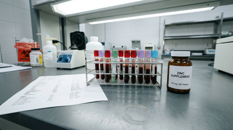
Hair Tissue Mineral Analysis (HTMA) provides insight into heavy metal excretion patterns over a 2-3 month window, revealing toxic burdens and mineral imbalances that blood tests often miss. Unlike serum testing, which reflects recent exposure, HTMA highlights longer-term trends alongside deficiencies in key minerals such as selenium, zinc, and magnesium that may further impair thyroid function.
At a Glance
- Mercury binds irreversibly to selenium, potentially depleting the deiodinase enzymes required for T4-to-T3 thyroid hormone conversion (Ralston and Raymond, 2010).
- Arsenic exposure is associated with thyroid peroxidase (TPO) inhibition and elevated TSH levels in multiple population studies.
- Cadmium has a biological half-life of 10-30 years and may accumulate in thyroid tissue at higher concentrations than surrounding organs.
- HTMA assesses mineral and heavy metal excretion over approximately 2-3 months, complementing single-point blood and urine tests.
- Standard TSH-only screening may not detect subclinical thyroid dysfunction driven by heavy metal-induced enzyme inhibition or receptor resistance.
- Selenium, zinc, magnesium, and iodine are key protective minerals that heavy metals can deplete or displace.
Understanding How Heavy Metals Affect Thyroid Function
The Thyroid-Heavy Metal Connection
The thyroid gland is particularly vulnerable to environmental toxins due to its role in concentrating iodine via the sodium-iodide symporter (NIS) and its high metabolic activity. Heavy metals may disrupt thyroid function through multiple mechanisms, including enzyme inhibition, nutrient depletion, oxidative damage, and immune dysregulation (1). Research published in Environmental Research by Zhang Y et al. (2019) systematically reviewed these associations across population-level studies.
A key challenge is that heavy metal-induced thyroid dysfunction often exists in a subclinical range. Symptoms may be significant while TSH remains within reference limits. This occurs because heavy metals can affect the hypothalamic-pituitary-thyroid (HPT) axis, deiodinase enzyme activity, receptor sensitivity, and cellular function, processes not fully captured by TSH alone (2). As Boas M et al. noted in Current Opinion in Endocrinology, Diabetes and Obesity (2012), environmental chemicals represent an underappreciated category of thyroid disruptors.
In our Adelaide-based functional medicine practice, HTMA frequently reveals heavy metal patterns in people with persistent thyroid symptoms, elevated thyroid antibodies, or poor response to thyroid medication, particularly when conventional thyroid panels appear “normal.”
Who this is for: People with fatigue, weight gain, brain fog, cold sensitivity, Hashimoto’s thyroiditis, Graves’ disease, known heavy metal exposure, or poor response to thyroid hormone replacement such as levothyroxine (T4) or liothyronine (T3).
Key insight: Heavy metal effects on thyroid function are often subclinical, meaning they may be detectable through HTMA and comprehensive thyroid testing before abnormalities appear on standard TSH-only screening.
Mercury: The Selenium Thief
Mercury is one of the most extensively studied thyroid-disrupting heavy metals, with research by Soldin OP et al. (2008) in Thyroid demonstrating direct interference with iodine metabolism and hormone synthesis.
| Source Category | Common Exposures |
|---|---|
| Dental | Amalgam fillings (approximately 50% mercury by weight) |
| Dietary | Contaminated fish, tuna, swordfish, shark, king mackerel |
| Environmental | Industrial emissions, coal combustion |
| Occupational | Mining, chlor-alkali plants, laboratory work |
How Mercury Disrupts Thyroid Function
1. Iodine uptake interference: Mercury competes with iodine at the sodium-iodide symporter (NIS), reducing iodine availability for thyroid hormone production. Studies suggest iodine uptake may be reduced by up to 50% in exposed populations (3).
2. Selenium depletion: Mercury binds tightly to selenium, forming irreversible mercury-selenide complexes that create a functional selenium deficiency even when dietary intake appears adequate (4). Ralston NVC and Raymond LJ demonstrated in Toxicology (2010) that selenium is essential for:
- Type 1, Type 2, and Type 3 deiodinase enzymes (T4-to-T3 conversion)
- Glutathione peroxidase (GPx), thyroid antioxidant protection
- Thyroid hormone receptor sensitivity
When selenium is depleted, T4-to-T3 conversion becomes impaired, leading to hypothyroid symptoms despite “normal” laboratory values.
3. Autoimmune activation: Mercury exposure has been associated with elevated thyroid antibodies, particularly anti-thyroglobulin antibodies (TgAb), suggesting a potential role in triggering autoimmune thyroid disease in genetically susceptible individuals. Havarinasab S et al. published supporting evidence in Environmental Health Perspectives (2007) (5).
Arsenic: The Enzyme Blocker
Arsenic exposure is associated with direct inhibition of thyroid peroxidase (TPO), the critical enzyme responsible for iodination and coupling reactions in thyroid hormone synthesis (6).
| Source Category | Common Exposures |
|---|---|
| Water | Contaminated groundwater (particularly in rural and bore-water regions) |
| Dietary | Rice products, certain seafood |
| Environmental | Industrial emissions, CCA-treated timber |
| Agricultural | Pesticides, herbicides |
How Arsenic Disrupts Thyroid Function
Thyroid peroxidase inhibition: Chowdhury S et al. demonstrated in the Journal of Trace Elements in Medicine and Biology (2014) that arsenic inhibits TPO, the enzyme required for iodination and thyroid hormone synthesis (6).
Oxidative damage: Arsenic generates reactive oxygen species (ROS) that damage thyroid follicular cells, contributing to inflammation and structural dysfunction. Mittal M et al. detailed this mechanism in Free Radical Biology and Medicine (2014) (7).
Compensatory TSH elevation: Impaired hormone synthesis may lead to increased TSH output via the HPT axis feedback loop. Wang Y et al. in Environmental Health (2018) and Paul DS et al. in Biological Trace Element Research (2015) demonstrated consistent associations between arsenic exposure and elevated TSH levels (8, 9).
Cadmium: The Accumulator
Cadmium has a biological half-life of 10-30 years in human tissue, allowing it to accumulate in thyroid tissue at concentrations higher than surrounding organs, as documented by Nordberg GF et al. in the Handbook on the Toxicology of Metals (2015) (11).
| Mechanism | Thyroid Effect | Key Finding |
|---|---|---|
| Hormone regulation disruption | Variable hypo- or hyperthyroid patterns | Chen X et al. (2013) documented cadmium accumulation in thyroid tissue (12) |
| Zinc displacement | Impaired thyroid hormone receptor activity | Ruhnke M et al. (2016) identified zinc-dependent receptor dysfunction (13) |
| Immune dysregulation | Increased Graves’ disease risk in smokers | Vestergaard P (2002) meta-analysis in European Journal of Endocrinology (14) |
Why Standard Thyroid Testing Misses These Patterns
Conventional thyroid screening typically measures TSH alone, which may fail to detect several categories of heavy metal-induced thyroid dysfunction.
| Limitation of TSH-Only Testing | What It Misses |
|---|---|
| No T4/T3 conversion assessment | Deiodinase enzyme inhibition from mercury or selenium depletion |
| No receptor sensitivity measure | Cadmium-induced zinc displacement at thyroid hormone receptors |
| No antibody screening | Early autoimmune thyroiditis (Hashimoto’s or Graves’ disease) |
| No toxicant exposure data | Underlying heavy metal burden driving subclinical dysfunction |
| Single time-point only | Chronic, low-level accumulation patterns visible on HTMA |
Hair Tissue Mineral Analysis (HTMA): A Window Into Heavy Metal Exposure
HTMA analyses a small sample of hair (typically from the first 3-4 cm of new growth at the nape of the neck) for toxic elements, essential minerals, and critical mineral ratios. Hair represents approximately 2-3 months of mineral excretion, providing longer-term insight than blood or urine testing (15). Seidel S et al. reviewed the clinical applications and limitations of hair mineral analysis in the Journal of Trace Elements in Medicine and Biology (2001).
| Testing Method | Time Window | Strengths | Limitations |
|---|---|---|---|
| Blood (serum) | Hours to days | Reflects acute exposure; widely available | Misses chronic accumulation patterns |
| Urine | Hours to days | Useful for provoked challenge testing | Reflects renal excretion, not tissue burden |
| HTMA (hair) | 2-3 months | Longer-term mineral and metal trends; non-invasive | Requires practitioner interpretation; not a standalone diagnostic |
Working With a Qualified Practitioner
HTMA interpretation and heavy metal detoxification should always be supervised by a qualified practitioner experienced in functional medicine and clinical nutrition. Improper protocols, such as aggressive chelation therapy without adequate mineral support, can worsen symptoms by redistributing metals into sensitive tissues including the brain and kidneys. A qualified practitioner can also assess underlying detoxification capacity, including factors such as methylation status, glutathione production, and metallothionein expression.
Next Steps
- Request HTMA testing: A Hair Tissue Mineral Analysis can reveal heavy metal excretion patterns and mineral imbalances that standard blood tests miss, providing a 2-3 month window of insight.
- Combine with comprehensive thyroid panels: Pair HTMA results with full thyroid testing, including free T4, free T3, reverse T3, and thyroid antibodies (TPOAb, TgAb), to identify subclinical dysfunction driven by toxic metal burden.
- Work with a qualified practitioner: Heavy metal detoxification requires supervised, safety-focused protocols to avoid redistributing metals into sensitive tissues.
Frequently Asked Questions
Key Insights
- Heavy metals can disrupt thyroid function without altering TSH, leading to under-recognised subclinical dysfunction.
- Mercury primarily affects thyroid health through selenium depletion and impaired T4-to-T3 conversion via deiodinase enzyme inhibition.
- Arsenic interferes with thyroid hormone synthesis by inhibiting thyroid peroxidase (TPO).
- Cadmium accumulates in thyroid tissue long term, altering hormone regulation and receptor signalling through zinc displacement.
- HTMA provides a 2-3 month view of mineral and metal patterns, complementing blood testing rather than replacing it.
Citable Takeaways
- Mercury competes with iodine at the sodium-iodide symporter (NIS) and may reduce iodine uptake by up to 50% in exposed populations, according to Soldin OP et al. in Thyroid (2008).
- Mercury-selenium binding creates irreversible complexes that deplete functional selenium, impairing T4-to-T3 conversion by deiodinase enzymes, as demonstrated by Ralston NVC and Raymond LJ in Toxicology (2010).
- Arsenic inhibits thyroid peroxidase (TPO) and is associated with elevated TSH levels in multiple population studies, including Wang Y et al. in Environmental Health (2018).
- Cadmium has a biological half-life of 10-30 years and accumulates preferentially in thyroid tissue, disrupting hormone regulation and zinc-dependent receptor activity, per Nordberg GF et al. (2015) and Ruhnke M et al. (2016).
- Hair Tissue Mineral Analysis (HTMA) assesses mineral and heavy metal excretion over approximately 2-3 months, providing a complementary window to single-point blood and urine testing, as reviewed by Seidel S et al. in the Journal of Trace Elements in Medicine and Biology (2001).
Uncover Hidden Thyroid Disruptors
If you are experiencing persistent thyroid symptoms despite “normal” results, heavy metal accumulation may be an overlooked contributor. At Elemental Health and Nutrition, we use HTMA and comprehensive thyroid testing to identify toxic metal patterns and mineral imbalances that standard screening misses, then design targeted, safety-focused protocols to support your recovery.
References
- Zhang Y et al. Environmental exposure to heavy metals and thyroid function: a systematic review. Environ Res. 2019 Mar;171:578-588. https://doi.org/10.1016/j.envres.2019.01.052
- Boas M et al. Environmental chemicals and thyroid function: an update. Curr Opin Endocrinol Diabetes Obes. 2012 Oct;19(5):385-391. https://doi.org/10.1097/MED.0b013e328357271a
- Soldin OP et al. Mercury and iodine interactions affecting thyroid function. Thyroid. 2008 May;18(5):521-527. https://doi.org/10.1089/thy.2007.0249
- Ralston NVC, Raymond LJ. Dietary selenium’s protective effects against methylmercury toxicity. Toxicology. 2010 Nov 28;278(1):112-123. https://doi.org/10.1016/j.tox.2010.04.015
- Havarinasab S et al. Mercury exposure and autoimmune response in the thyroid. Environ Health Perspect. 2007 Apr;115(4):569-574. https://doi.org/10.1289/ehp.9411
- Chowdhury S et al. Arsenic exposure and thyroid peroxidase inhibition. J Trace Elem Med Biol. 2014 Oct;28(4):359-365. https://doi.org/10.1016/j.jtemb.2014.07.003
- Mittal M et al. Oxidative stress and arsenic-induced thyroid dysfunction. Free Radic Biol Med. 2014 Aug;73:302-309. https://doi.org/10.1016/j.freeradbiomed.2014.05.026
- Wang Y et al. Association between arsenic exposure and serum TSH levels in adults. Environ Health. 2018 Nov 22;17:81. https://doi.org/10.1186/s12940-018-0428-5
- Paul DS et al. Chronic arsenic exposure and thyroid hormone alterations. Biol Trace Elem Res. 2015 Apr;164(2):183-190. https://doi.org/10.1007/s12011-014-0222-3
- Nishijo M et al. Thyroid hormone changes in populations exposed to environmental metals. Environ Res. 2017 Feb;153:157-164. https://doi.org/10.1016/j.envres.2016.11.021
- Nordberg GF et al. Cadmium. In: Handbook on the Toxicology of Metals. 4th ed. Academic Press; 2015:445-486.
- Chen X et al. Cadmium accumulation in thyroid tissue and endocrine disruption. J Appl Toxicol. 2013 Oct;33(10):940-946. https://doi.org/10.1002/jat.2875
- Ruhnke M et al. Zinc displacement and thyroid hormone receptor dysfunction. Mol Cell Endocrinol. 2016 Nov 15;437:193-202. https://doi.org/10.1016/j.mce.2016.08.019
- Vestergaard P. Smoking and thyroid disorders: a meta-analysis. Eur J Endocrinol. 2002 Feb;146(2):153-161. https://doi.org/10.1530/eje.0.1460153
- Seidel S et al. Hair mineral analysis: clinical interpretation and limitations. J Trace Elem Med Biol. 2001;15(1):1-9. https://doi.org/10.1016/S0946-672X(01)80001-8